
Figure 8.
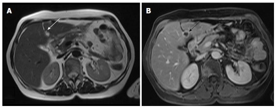
Cervical cancer, stage IVB. A: Axial T2-weighted image shows a hyperintense lesion within the falciform ligament (arrow); B: Corresponding axial T1-weighted image after intravenous injection of paramagnetic contrast shows the necrotic nature of the lesion (arrow). Cavitating metastatic lesions are usually indicative of cervical carcinomas of squamous cell origin.