Figure 3.

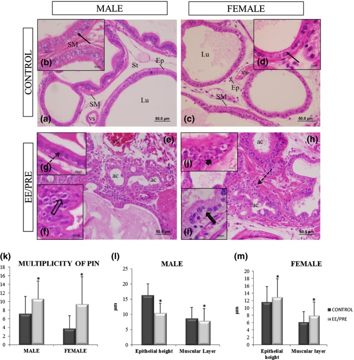
Histological sections of the ventral male prostate and female prostate from senile gerbils stained by haematoxylin and eosin (HE). Control group: (a, c) Prostatic acini surrounded by a simple prismatic epithelium (Ep) with the presence of luminal region (Lu), smooth muscle layer (SM) and stroma (St) with vessels (vs). (b, d) Detail of the epithelial height (arrows). EE/PRE group: (e, h) Acinar atrophy (ac). (f, j) Presence of prostatic intraepithelial neoplasia and atypical nuclei (broad arrow and arrowhead). (g) Decrease in epithelial height in the ventral male prostate (dashed arrow). (i) An increase in epithelial height (broad arrow) has been observed in the female prostate. (k) Multiplicity (specific number) of prostatic neoplasia intraepithelial (PIN) for each experimental senile male and female. (l, m) Morphometric of epithelial heights and thicknesses of the muscle layers of the ventral male prostate and female prostate from senile gerbils of the experimental groups. Values of multiplicity of PIN and morphometry are expressed as the mean ± SD. EE/PRE: exposure to ethinylestradiol during the prenatal period. *Significant difference between the groups with P ≤ 0.05.
