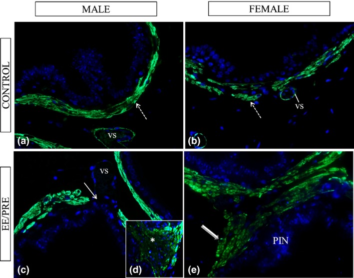
Figure 5.

Histological sections of the ventral male prostates and female prostates from senile gerbils subjected to immunofluorescence for α‐actin of smooth muscle. Control group: (a and b) Immunoreactivity in the prostatic muscle layer (dashed arrows) and in the vessels (vs). EE/PRE group: (c–e) Region with prostatic buds noted absence of this immunoreactivity (arrow). Observed an increase in the α‐actin immunolocalization in lesions in the stroma (*) and in the regions with PIN (large arrow).
