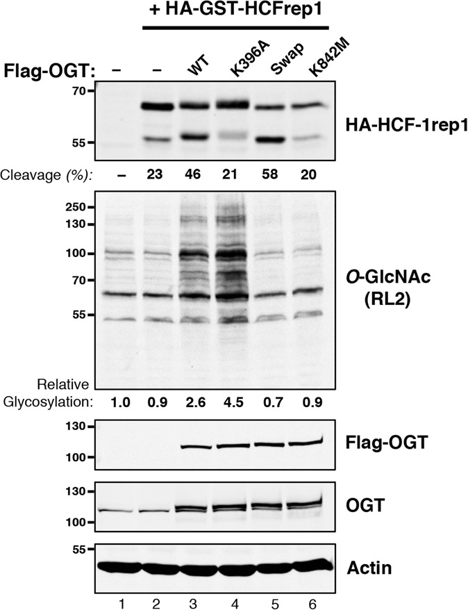
Figure 6.

In vivo glycosylation and HCF-1rep1 cleavage properties of activity-selective OGT mutants. HEK293 cells were untransfected (lane 1), transfected with HA-GST–HCF-1rep1 vector alone (lane 2), cotransfected with wild-type (lane 3) or mutant (lanes 4–6) Flag-OGT expression vectors as described in the Materials and Methods. (Top panel) HA-GST–HCF-1rep1 cleavage was detected using anti-HA antibody and quantified as described in the Materials and Methods. Endogenous HEK293 protein O-GlcNAcylation was visualized with anti-O-GlcNAc RL2 antibody. Levels of ectopic recombinant Flag-OGT were detected alone or in combination with endogenous OGT with anti-Flag or anti-OGT antibody, respectively. (Bottom panel) An anti-actin blot is shown as a loading control. To quantitate O-GlcNAcylation of endogenous proteins, the total combined intensity of O-GlcNAcylated proteins in the untransfected HEK29 cells was assigned an arbitrary value of 1. The relative intensities of O-GlcNAcylation in each transfected sample were then calculated as a ratio between its combined protein O-GlcNAcylation intensity over that of the untransfected lysate. The quantitation indicated is for the experiment shown. Similar results were obtained in three additional separate experiments.
