
Figure 3. Binding of wild-type IGFBP7 and IGFBP7 fragments to insulin shown by western blot.
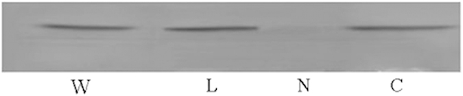
Up to 20 μl insulin (1 μg/μl) was subjected to SDS–PAGE and then electroblotted on to nitrocellulose membranes. The membranes were then incubated with wild-type IGFBP7 or fragments. After washing, the membranes were incubated with anti-His antibody and then incubated in fluorescence-labeled secondary antibodies. The bands were detected with the imaging system and quantified. Lanes from left to right, wild-type IGFBP7, IGFBP7-L fragment (172–244), IGFBP7-N fragment (1–171), IGFBP7-C fragment (245–282).