
Figure 4. Ligand dot blot of IGFBP7 mutants with insulin and IGFBP7 antibody. 2.5 μl (0.09 mg/ml) wild-type IGFBP7 and IGFBP7 mutants were dotted directly on to the nitrocellulose membrane.
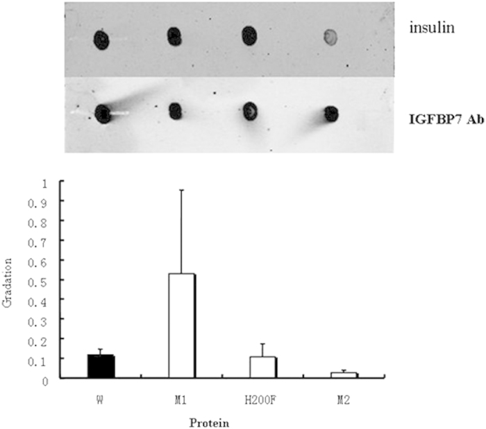
After blocking, the membrane was incubated at 4 °C overnight with 1 μg/ml insulin. Membranes were washed and incubated with insulin antibody (upper panel). Lanes from left to right, wild-type IGFBP7, R198E mutant, H200F mutant, R198I-H200F mutant. Membranes were incubated with IGFBP7 antibody (lower panel), showing the presence of approximately equal amounts of protein. All blots shown are representative of at least three separate experiments, with error bars representing SD. The graph shows densitometric analysis of dots.