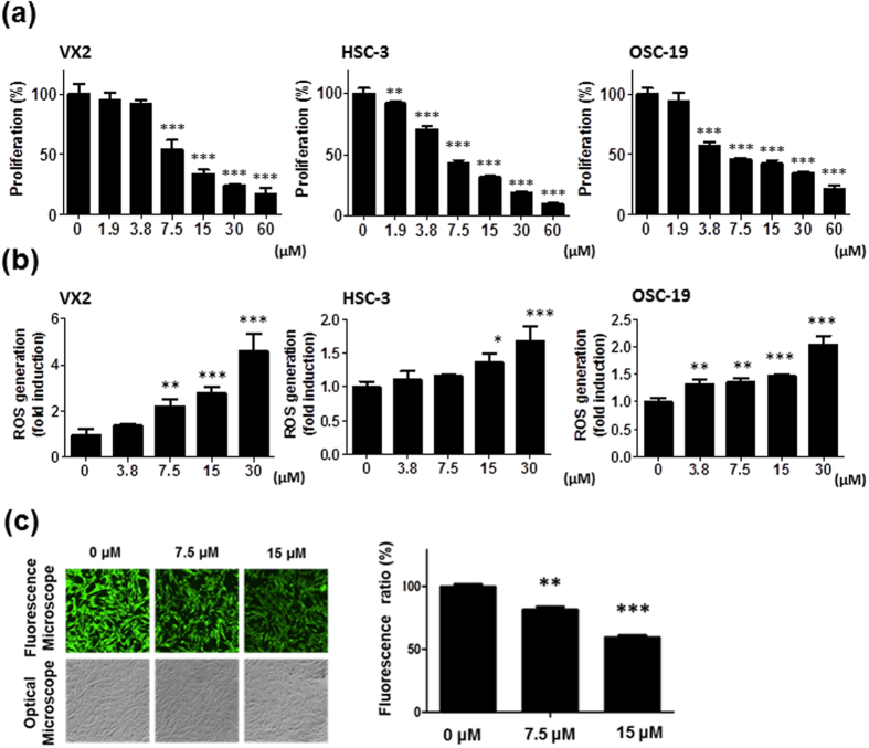
Figure 2. Fe(Salen) nanoparticles inhibit cell proliferation, promote ROS generation, and are taken up by cells.
(a) Effect of Fe(Salen) on proliferation of various cancer cells. XTT cell proliferation assays were performed with human and rabbit tongue cancer cells: VX2 rabbit squamous cell carcinoma, HSC-3 human squamous cell carcinoma, and OSC-19 human squamous cell carcinoma (n = 4). The IC50 values were similar among cell types and were approximately 7.5 μM. (b) Effect of Fe(Salen) on ROS production in various cells. Fe(Salen) nanoparticles generated ROS in a concentration-dependent manner (n = 4, **p < 0.01, ***p < 0.001). (c) Representative fluorescence pictures of calcein using a fluorescence microscope and optical microscope. Ratios of calcein fluorescence are shown below (n = 4, **p < 0.01, ***p < 0.001 vs. control). Note that cellular fluorescence was decreased in the presence of Fe(Salen) nanoparticles.

