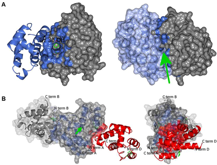
Figure 2. The protein-protein interface in the crystal structure of MvicOBP3 (A) and NribOBP3 (B).
The green arrows indicate the binding pockets. Tyr30 is shown in green. (A) Left: the interface of chain A (grey surface with interface residues in blue) and chain B (blue ribbon) with TyrB30 (green). Right: the view of horizontal 90 degree rotation around the pocket entrance. Chain B is in a light blue surface. (B) Left: chains B (grey), A (blue) and D (red) in the filament. The surfaces of A and B are grey with the interface coloured by the interacting chain. Right: the end view of three fold rotation along the filament. The three green Tyr30s indicate the three fold.

