Figure 4.
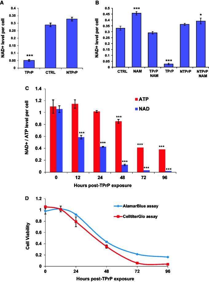
TPrP induces depletion of intracellular NAD+ and ATP. (A) TPrP at 5 µg/ml for 3 days induces intracellular NAD+ depletion, but not non-toxic PrP at 10 µg/ml. CTRL = untreated cells. Statistical differences are shown compared to CTRL. The experiment was done more than four times with duplicate samples; standard deviations are shown. (B) TPrP-induced NAD+ depletion is corrected by the addition of nicotinamide (100 µg/ml). Cells were treated for 3 days with TPrP at 5 μg/ml or non-toxic PrP at 5 μg/ml. Statistical differences are shown compared to CTRL. The experiment was done twice with duplicate samples; standard deviations are shown. (C) TPrP induces a time-dependent decrease of intracellular NAD+ (up to 200-fold) starting 12 h after TPrP exposure, correlating with a decrease of intracellular ATP (up to ∼3-fold) starting 36 h later. Statistical differences are shown compared to the zero time point. The experiment was done three times in triplicate wells; standard deviations are shown. (D) Time course of TPrP-induced toxicity measured by CellTiterGlo® and AlamarBlue® assays. (C and D) TPrP was used at a dose of 5 µg/ml. The experiment was done twice with triplicate samples; standard deviations are shown. *P < 0.05; ***P < 0.001.

