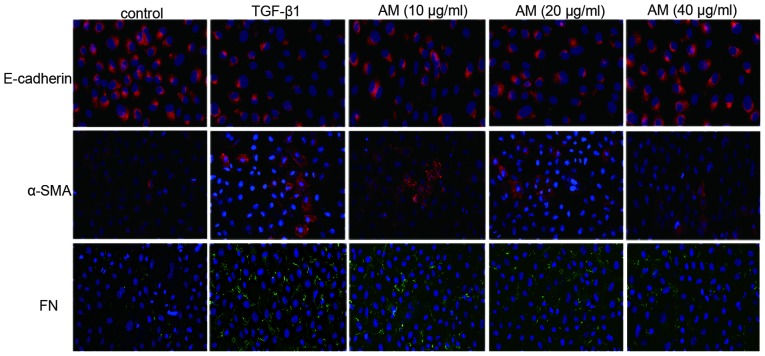
Figure 3.
Murine renal proximal tubule NRK-52E cells were stained with E-cadherin, α-SMA, and FN to determine epithelial-mesenchymal transition (magnification, ×200). In the control group, no α-SMA or FN-positive cells were detected, whereas numerous E-cadherin-positive cells were detected. E-cadherin expression decreased in the TGF-β1 group, whereas the number of α-SMA and FN-positive cells increased, as compared with the control group. Groups treated with 10, 20, and 40 µg/ml AM exhibited reverse effects, as compared with the TGF-β1 group. SMA, smooth muscle actin; FN, fibronectin; TGF, tumour growth factor; AM, Astragalus membranaceus.

