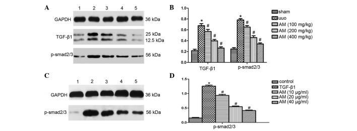
Figure 5.
Smad-dependent TGF-β1 signaling in UUO mice and murine renal proximal tubule NRK-52E cells, as detected by western blotting and quantified using densitometric analysis. (A) Representative western blotting and (B) semiquantitative histograms of TGF-β1 and p-Smad2/3 expression levels in UUO mice. Data are presented as the mean ± standard deviation. *P<0.05, vs. the sham group; #P<0.05, vs. the UUO group. (C) Western blotting and (D) semiquantitative histograms of p-Smad2/3 expression in the groups treated with TGF-β1 alone and TGF-β1 with AM in NRK-52E cells. Data are presented as the mean ± standard deviation. *P<0.05, vs. the control group; #P<0.05, vs. the TGF-β1 group. TGF, tumour growth factor; UUO, unilateral ureteral obstruction; p phosphorylated; AM, Astragalus membranaceus.

