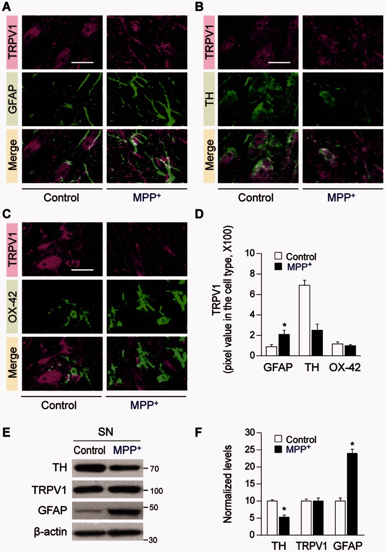
Figure 3.
TRPV1 expression in the substantia nigra of MPP+-lesioned rat. The rats were given a unilateral medial forebrain bundle (MFB) injection of MPP+ and brain tissues were processed for immunohistochemical and western blot analysis at 1 week post MPP+. (A–D) Fluorescence images of TRPV1 (magenta; A) and GFAP (green; A), or TRPV1 (magenta; B) and TH (green; B) or TRPV1 (magenta; C) and OX-42 (green; C) and both images are merged (yellow; A–C) in the SNpc of MPP+-lesioned rat brain. (D) Quantification of TRPV1 expression in each cell type, *P < 0.01, **P < 0.001, significantly different from control. (E and F) Western blot analysis of TH, TRPV1 and GFAP (E), and quantification (F) in the SN of MPP+-lesioned rat brain, *P < 0.001, significantly different from control. Scale bars = 20 µm. Mean ± SEM; D, n = 6–7; F, n = 4–6. ANOVA and Student-Newman-Keuls analysis.

