Figure 6.
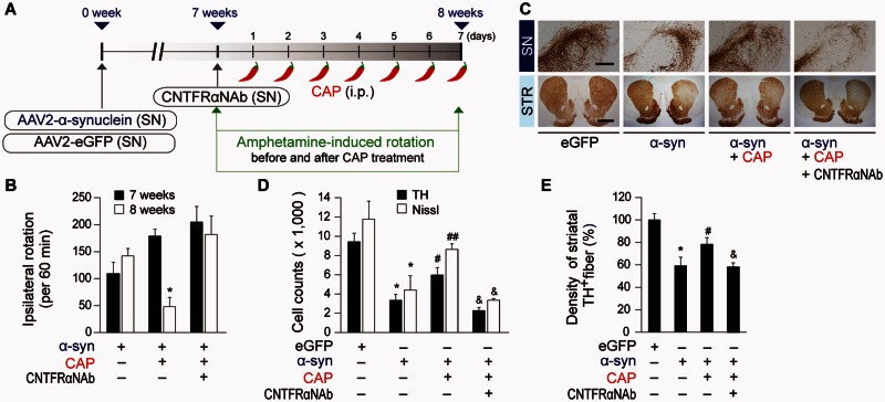
Capsaicin prevents α-synuclein-induced degeneration of dopamine neurons in vivo. (A) Diagram of the experimental design. Rats were given a unilateral injection of AAV2-α-synuclein (α-syn) or AAV2-eGFP (eGFP, control) into the rat SN and injection of CNTFRα neutralizing antibody (CNTFRαNAb) into the SN at 7 weeks post AAV2-α-synuclein. All rats received capsaicin (CAP; 1 mg/kg, intraperitoneally) or vehicle at 7 weeks and 1 day after injection of AAV2 and a continuous single injection per day for 7 days. Rats were transcardially perfused after the last rotation experiment. (B) Cumulative amphetamine-induced ipsilateral rotations, *P < 0.01, significantly different from 7weeks α-synuclein. (C) Photomicrographs of TH+ cells in the SN and TH+ fibres in the striatum (STR). (D) Number of TH+ or Nissl+ cells in the SNpc, *P < 0.001, significantly different from eGFP, #P < 0.05, ##P < 0.01, significantly different from α-synuclein, &P < 0.01, significantly different from α-synuclein + capsaicin. (E) Optical density of TH+fibres in the striatum, *P < 0.01, significantly different from eGFP, #P < 0.05, significantly different from α-synuclein, &P < 0.05, significantly different from α-synuclein + capsaicin. Scale bars: 400 µm (SN), 2 mm (STR). Mean ± SEM; B–E, n = 4–10 in each group. ANOVA and Student-Newman-Keuls analysis.

