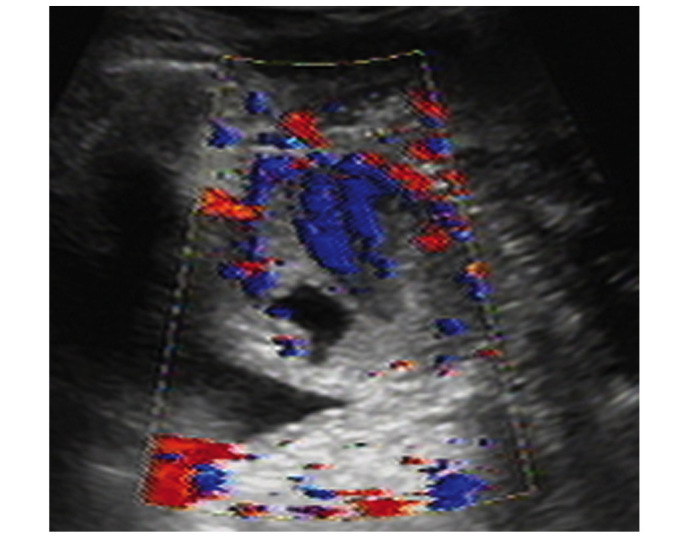
Figure 2.

Transvaginal ultrasound demonstrated a hypoechoic circumscribed but not well-demarcated mass. Color Doppler flow imaging revealed predominantly low resistance type arterial blood flow between the paries anterior vagina and posterior bladder wall. The size of the mass was 4.9×3.6×3.0 cm.
