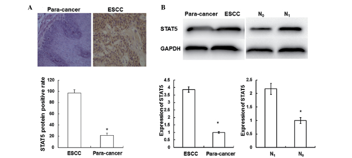
Figure 2.
Immunohistochemical staining and western blotting were performed to investigate the expression of STAT5 in ESCC tissues. Experiments were repeated ≥3 times. (A) Expression of STAT5 in ESCC tissues and adjacent normal tissues was detected using immunohistochemical staining. Cells with brown staining were defined as STAT5-positive (magnification, ×200). *P<0.05 vs. ESCC. (B) The expression of STAT5 in ESCC tissues, adjacent normal tissues and tissues from patients with or without lymph node metastasis was detected using western blotting. *P<0.05 for para-cancer vs. ESCC or for N0 vs. N1. GAPDH was used as a loading control. ESCC, esophageal squamous cell carcinoma; STAT5, signal transducer and activator of transcription; GAPDH, glyceraldehyde 3-phosphate dehydrogenase; N1, with lymph node metastasis; N0, without lymph node metastasis.

