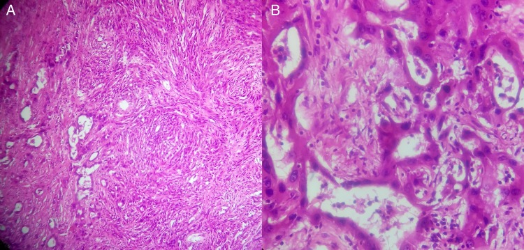
Figure 2.
(A) HPE depicting biphasic morphology with epithelial cells forming tubules and sheets, and a sarcomatoid component having numerous spindle cells with stromal invasion and areas of necrosis (H&E ×100). (B) Note the cuboidal cells lining the tubules, with high nucleocytoplasmic ratio, mitotic figures (3/50 high-power field) and atypical mitotic figures (H&E ×400).

