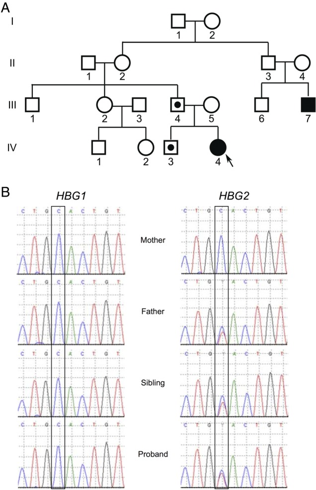
Figure 1.

The proband has a paternally inherited mutation in HGB2 that results in Hb FM-Fort Ripley. (A) Family pedigree with IV-4 representing the proband. Black filled symbols represent individuals who presented with neonatal cyanosis. The father (III-4) and sibling (IV-3) of the proband have red blood cell indices suggestive of an α-thalassaemia trait, which is indicated by a star. (B) Chromatograms from the indicated individuals showing HBG1 and HBG2. Long-range PCR using primers from Crowley et al was performed followed by dideoxy sequencing.
