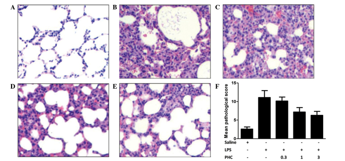
Figure 1.
Histology and pathological scoring of the left lung tissue samples in LPS-treated rats with or without PHC treatment. Rats were treated with 8 mg LPS/kg body weight for 1 min prior to treatment with the indicated concentrations of PHC (mg/kg body weight). After 6 h of treatment, the left lung tissue samples were harvested and stained with hematoxylin and eosin. The tissue sections were analyzed by light microscopy (magnification, ×400). (A) Control group; (B) LPS-only group; (C) 0.3, (D) 1 and (E) 3 mg PHC/kg body weight + LPS groups. (F) Pathology of the left lung tissue samples was scored using Smith-scoring. Data are presented as the mean ± standard deviation. *P<0.05 vs. the saline control; #P<0.05 vs. the LPS-treated groups. TNF-α, tumor necrosis factor-α; IL-6, interleukin-6; LPS, lipopolysaccharide; PHC, penehyclidine.

