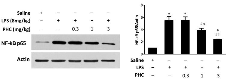
Figure 4.
NF-κB p65 expression in the left lung tissue samples of LPS-treated rats. Rats were treated with 8 mg/kg body weight LPS for 1 min prior to treatment with the indicated concentrations of PHC (mg/kg body weight) for 6 h. Western blot analysis was performed using NF-κB p65 and β-actin, which was used as the internal control. Treatment with 1 and 3 mg/kg LPS significantly attenuated the LPS-induced upregulation of NF-κB p65 expression. *P<0.05 vs. the saline control #P<0.05 and ##P<0.01 vs. the LPS-treated groups. NF-κB, nuclear factor-κB; PHC, penehyclidine; TLR2/4, toll-like receptor 2/4.

