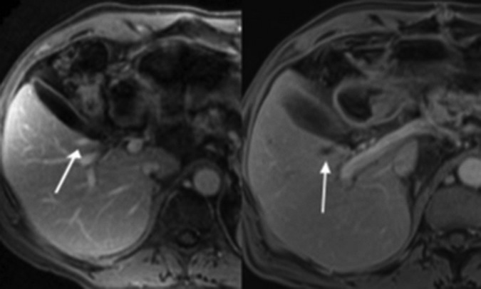
Figure 2.
(A) Preablation MRI axial T1-weighted volumetric interpolated breath-hold examination (VIBE) postgadolinium portal venous phase images show normal enhancement of the right inferior hepatic vein branch. (B) Postablation the vessel is expanded and lacks enhancement, consistent with thrombosis.

