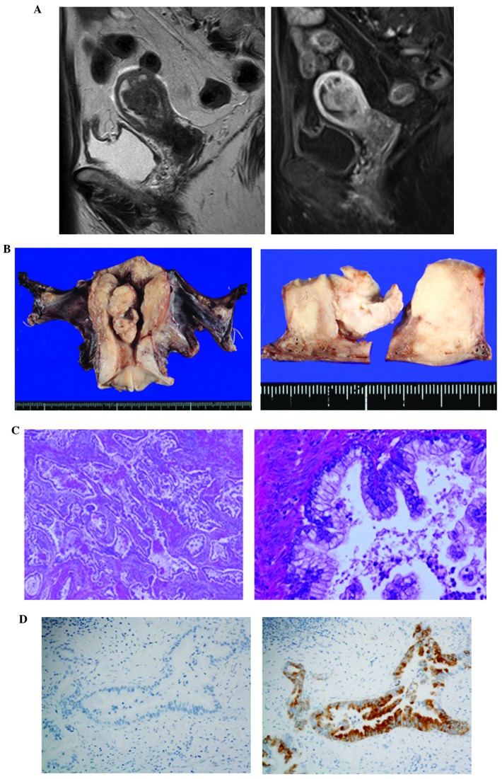
Figure 1.
Magnetic resonance imaging (MRI) and pathological findings in case 1. (A) T2-weighted MRI scan showing a tumor with intermediate intensity (left panel). Enhanced T1-weighted images showing that the tumor invaded over half the thickness of the myometrium (right panel). (B) Macroscopically, the tumor was whitish, sized 5.4 cm, arising from the left wall and invading over half of the myometrium. (C) The microscopic findings included a diffusely infiltrating mucinous adenocarcinoma with well-formed glands, aggressively invading the myometrium, along with a desmoplastic stromal reaction; (left panel) magnification, ×100; right panel magnification, ×400. (D) On immunohistochemistry, the tumor cells were positive for MUC6 (right panel), but negative for HIK1083 (left panel); magnification, ×400.

