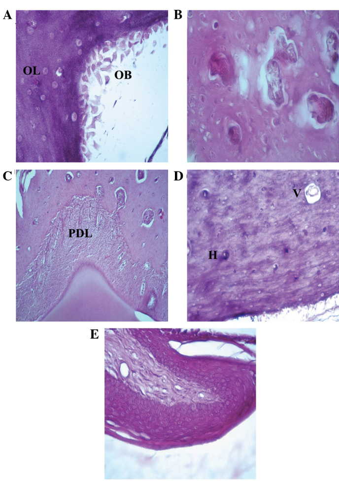
Figure 2.

Hematoxilin and eosin staining of the rat mandibular bone and gingival epithelium after 7–15 days of zoledronate treatment (0.1 mg/kg). (A) Osteoblasts at the rim side of the cortical bone and full osteocytic lacunae were observed (magnification, ×40). Hematoxylin and eosin staining also demonstrated the presence of (B) full Howship's lacunae (magnification, ×40), (C) normal periodontal ligament between the tooth root and the bone (magnification, ×10) and (D) full Volkman's and Haversian systems (magnification, ×20). (E) Normal gingival epithelium structure (magnification, ×20). OB, osteoblast; OL, osteocytic lacunae; PDL, periodontal ligament; V, Volkman's systems; H, Haversian systems.
