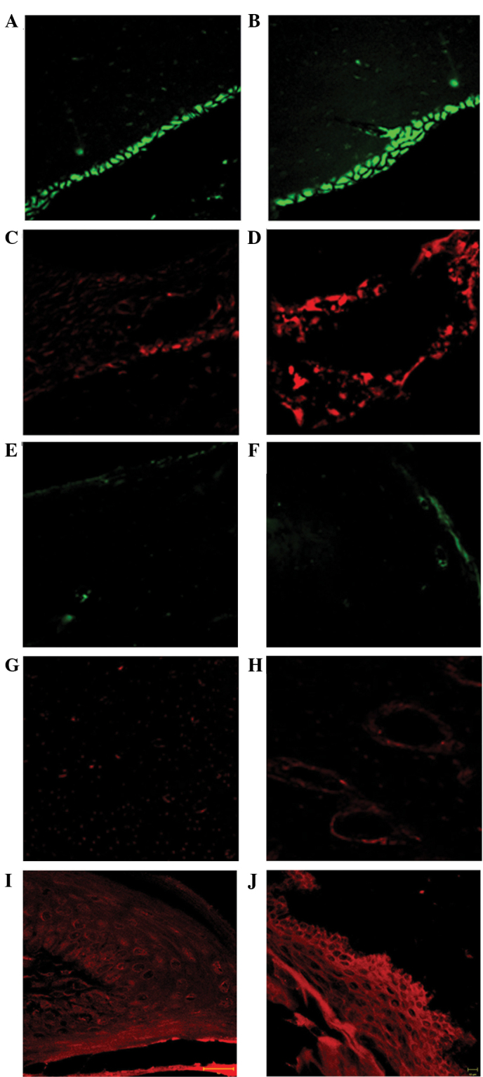
Figure 4.

Immunofluorescence staining of the rat mandibular bone and gingival epithelium following 7–15 and 30–45 days of zoledronate treatment (0.1 mg/kg). In the 7–15 day group, (A and B) osteoblasts were present at the mineralization side, as determined by osteocalcin labeling (green), as well as (C and D) the presence of osteoclasts, labeled by RANK (red), in the Howship's lacunae. In the 30–45 days group a near total absence of the tested proteins, (E and F) osteocalcin and (G and H) RANK was observed. (E) In all layers of the gingival epithelium a uniform α-sarcoglycan pattern of fluorescence (red) was observed in the (I) 7–15 and (J) 30–45 day groups. Magnifications are ×40 for A-H, ×63 for I and ×20 for J. RANK, receptor activator of nuclear factor-κB.
