Fig. 6.

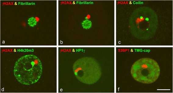
a–e Confocal microscopy images of SGN nuclei co-stained for γH2AX in combination with fibrillarin (a, b), coilin (c), histone H4K20me3 (d) and HP1γ (e). They illustrate the direct association of PDDF with a triad of structures: the nucleolus (a, c, e), Cajal bodies, immunolabeled for fibrillarin (b) or coilin (c), and perinucleolar heterochromatin clumps immunolabled for the tri-methylated histone H4K20me3 (d) or HP1γ (e). f Double immunolabeling for 53BP1 and TMG-cap illustrate the distribution of the nuclear speckles of splicing factors labelled with the anti-TMG-cap antibody and the close association of a PDDF with a speckle in the vicinity of the nucleolus. 45d post-IR. Scale bar = 5 μm
