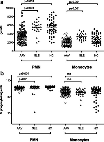
Fig. 4.

Decreased phagocytosis in anti-neutrophil cytoplasmic antibodies associated vasculitides (AAV). To evaluate further the function of phagocytes in AAV, the capacity to phagocytose opsonized E. coli was investigated in polymorphonuclear leukocytes (PMN) and monocytes from patients with AAV (n = 84), patients with systemic lupus erythematosus (n = 26), and healthy controls (HC) (n = 54). a Amount of phagocytosed E. coli bacteria shown as geometric mean fluorescence intensity (geoMFI). b Percentage of phagocytosing cells. Differences between healthy controls, patients with SLE, and patients with AAV were calculated using the Kruskal-Wallis test with Dunn’s multiple comparison test, and the following p values were obtained for all three groups: geoMFI of PMN, p < 0.0001; geoMFI of monocytes, p < 0.0001 and % phagocytosing cells of PMN, p < 0.0001; and % phagocytosing cells of monocytes, p = 0.4091. P values presented in the figure are for comparison between two groups
