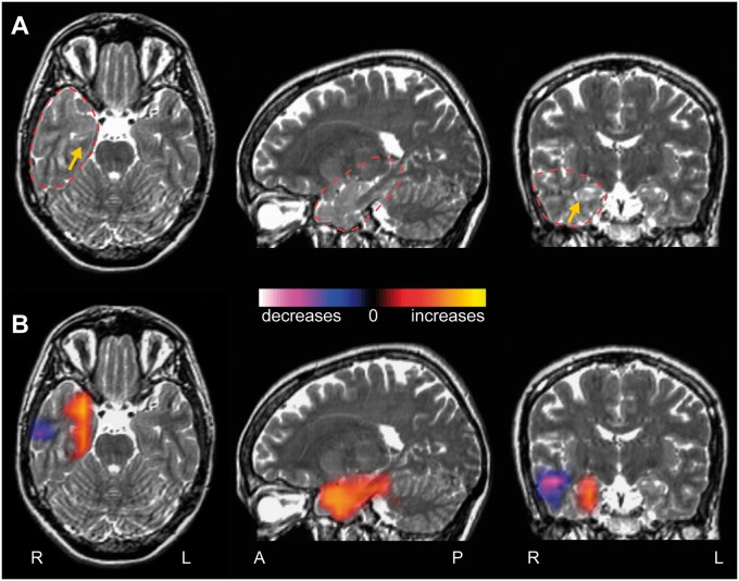
Figure 5.
Example patient with increased connectivity at the resection region. (A) T2-weighted MRI images from a 34-year-old right-handed female suffering from intractable focal dyscognitive and secondarily-generalized seizures for 21 years. The right anterior hippocampus shows slight T2 signal hyperintensity and blurred cytoarchitecture compared to the left side, suggestive of mesial temporal sclerosis (yellow arrow). Dashed red line represents region used for connectivity analysis. (B) A regional functional connectivity map (R-image) shows increased connectivity between the mesial temporal lobe and the rest of the brain, compared to corresponding voxels in the contralateral hemisphere. A smaller area of decreased connectivity is also seen in the lateral temporal cortex.

