. 2015 Mar 16;28(5):707–719. doi: 10.1080/08912963.2015.1020423

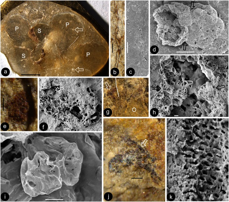
Figure 6 . (Colour online) Stamens of Euanthus panii gen. et sp. nov. Stereomicroscopy and SEM. (a) Nitro cellulose replica of the specimen in Figure 4(b), showing the positions of two anthers (white arrows) relative to the sepals (S) and petals (P). The white line marks the position of the possible filament shown in (b) and (c). Bar = 2 mm. (b, c) A possible filament on the replica, marked with a white line in (a). Stereomicroscopy (b) and SEM (c). Bar = 0.1 mm. (d) The anther marked by a lower arrow in (a) shows the constriction (arrows) between the left and right halves of the anther. The left half is broken, and its internal details are visible. Bar = 0.1 mm. (e) Dark organic material of the anther, marked by a blue arrow in Figure 4(b) and a lower white arrow in (a). Bar = 0.1 mm. (f) The anther marked by the upper arrow in (a), showing a broken anther with possible in situ pollen grains (arrow). Bar = 20 μm. (g) Details of the portion marked by the black arrow in Figure 4(e), showing a possible filament stub (arrow) beside the hairy style (to the right of white line) and the ovarian cavity (O). Bar = 0.1 mm. (h) Details of (d), showing two confluent pollen sacs in the anther (arrows) and its cellular details. Bar = 10 μm. (i) Detailed view of the possible in situ pollen grains in the anther shown in (f). Bar = 5 μm. (j) Organic material preserved in the flower, enlarged from the region marked by the white arrow in Figure 4(e). Bar = 0.1 mm. (k) Pitting on a vascular element, enlarged from the arrowed region in (j). Bar = 2 μm.

Figure 6