. 2015 Mar 16;28(5):707–719. doi: 10.1080/08912963.2015.1020423

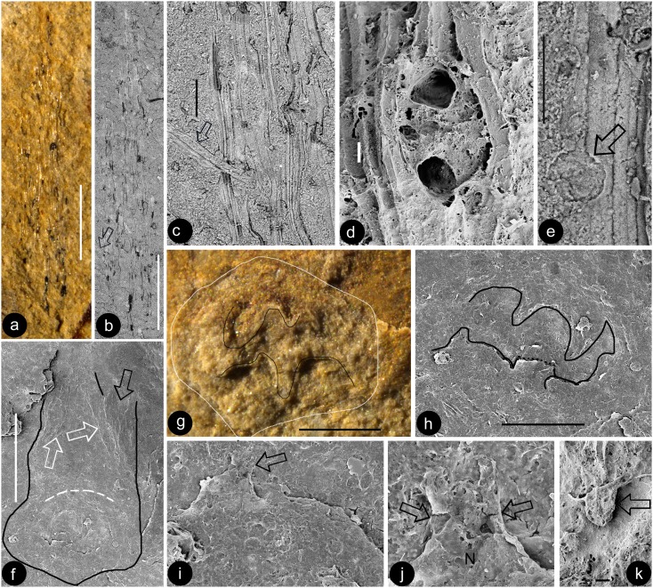
Figure 7 . (Colour online) Gynoecium of Euanthus panii gen. et sp. nov. Stereomicroscopy and SEM. (a, b) The distal style with hairs, viewed under SEM (a) and stereomicroscope (b). Bar = 1 mm. (c). A hair (arrow) branching off from the style, enlarged from the arrowed region in (b). Bar = 0.1 mm. (d) A possible secretory structure in the style. Bar = 10 μm. (e) Cells in the style with straight cell walls. Note a scar (arrow) left by a fallen-off hair. Bar = 50 μm. (f) The basal portion style and ovary (outlined). Note the branching-off possible filament stub (black arrow) and inner wall (white arrows) of the ovary. Refer to Figure 4(e). Bar = 1 mm. (g, h) Detailed view of the same receptacle and ovary, under stereomicroscope and SEM. Note the pentamerous outline of the receptacle (white line) and protrusions (black lines) on the inner wall of the ovary. Bar = 0.5 mm. (i) The ovule enlarged from (h), with a micropyle (arrow). Bar = 50 μm. (j) Details of the micropyle in (i). Note that there is only one layer of integument (arrows) covering the nucellus (n). Refer to Figure 8(b). Bar = 20 μm. (k) One of the papillae on the inner ovarian wall. Bar = 10 μm.

Figure 7