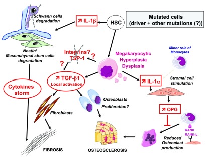
Figure 2. Role of microenvironment in the development of myelofibrosis.
Mutated dysplastic megakaryocytes (MKs) are responsible for the myelofibrosis and osteoclerosis by inducing the release of (i) non-activated transforming growth factor β1 (TGFβ1), which is activated in the bone marrow environment by a so far uncharacterized mechanism, possibly via integrins and matrix such as fibronectin and thrombospondin (TSP). Fibrosis begins around MKs associated with the proliferation of fibroblasts and eventually osteoblasts, (ii) interleukin-1α (IL-1α) is released and induces osteoprotegerin (OPG) by t stromal cells, a decoy receptor that blocks osteoclast production. Mutated hematopoietic stem cells (HSCs) induce the increase in IL-1α and the subsequent degradation of Schwann cells and mesenchymal stem cells, leading to fibrosis and osteosclerosis through cytokine storm and providing a favorable environment for the hematopoietic clone.

