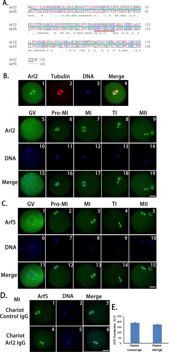
Figure 2. Peptide nanoparticle-mediated antibody transfection can specifically inhibit target protein in mouse oocytes.
(A) Protein sequence alignment of Arl2 and Arf5. Blue-underlined Arl2 sequence (2–182 AA) are antigen regions for the anti-Arl2 antibody, red-underlined Arf5 sequence (96–106 AA) are antigen regions for the anti-Arf5 antibody. B1–B4, co-localization of Arl2 and microtubules; B5–B19, Arl2 immunolocalization in mouse oocytes at each meiotic stage. Respectively, by column, B5, B10, B15, at GV stage; B6, B11, B16, Pro-MI; B7, B12, B17, MI; B8, B13, B18, TI; B9, B14, B19, MII; by line, B5–B9, Arl2 in green; B10–B14, DNA in blue; B15–B19, merge of Arl2 and DNA. (C) Arf5 immunolocalization in mouse oocytes at each meiotic stage. C1, C6, C11, at GV stage; C2, C7, C12, Pro-MI; C3, C8, C13, MI; C4, C9, C14, TI; C5, C10, C15, MII; by line, C1–C5, Arl2 in green; C6–C10, DNA in blue; C11–C15, merge of Arf5 and DNA. (D) Immunostaining of Arf5 at MI stage in control IgG (D1–D3) or anti-Arl2 antibody (D4–D6) transfection group. (E) Quantification of Arf5 fluorescence at MI stage in control IgG (left) or anti-Arl2 antibody (right) transfection oocytes. Scale bar, 20 μm.

