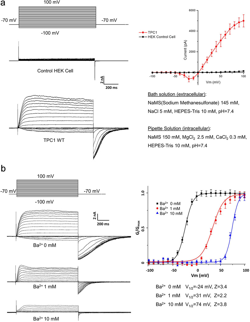
Extended Data Figure 2. Voltage activation and Ba2+ modulation of AtTPC1 over-expressed in HEK cell.
a, Voltage dependent activation of wild-type AtTPC1. Channel currents were recorded using patch clamp in the whole-cell configuration. The membrane was stepped from holding potential (−70 mV) to various testing potentials and then returned to the holding potential. The I–V curve was plotted using the steady peak current against the voltage. The peak tail currents were recorded to generate the G-V curves for voltage activation analysis. b, Extracellular Ba2+ inhibition of AtTPC1. The intracellular solution (pipette) contains 300 µM Ca2+ necessary for channel activation.

