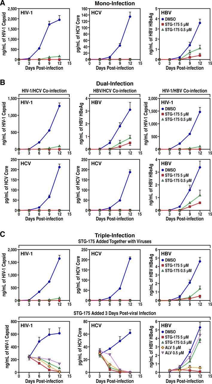
Fig 7. Mono- and co-infection analyses.
A. Mono-infection. Left panel. Activated PBMCs were exposed to HIV-1 JR-CSF (TCID50 of 103/mL) together with or without STG-175 for 3 h, washed and replication monitored by HIV-1 p24 ELISA for 12 days. Middle panel. Huh7.5.1 cells were exposed to HCV JFH-1 (TCID50 of 103/mL) for 3 h, washed and replication monitored by HCV core ELISA for 12 days. Right panel. NTCP-Huh7 cells were exposed to HBV AD38 (TCID50 of 106/mL) for 3 h, washed and replication monitored by HBV HBeAg ELISA for 12 days. B. Dual-infection. Left panels. Same as A, except that mixed PBMCs and Huh7.5.1 cells were exposed to both HIV-1 and HCV. Middle panels. Same as A, except that mixed NTCP-Huh7 and Huh7.5.1 cells were exposed to both HBV and HCV. Right panels. C. Same as A, except that mixed PBMC and NTCP-Huh7 cells were exposed to both HIV-1 and HBV. C. Triple-infection. Same as A, except that mixed PBMCs, NTCP-Huh7 and Huh7.5.1 cells were exposed to HIV-1, HBV and HCV altogether. DMSO, STG-175 or ALV was added to cells either together with viruses (top panels) or 3 days after virus addition and then every 3 days (bottom panels).

