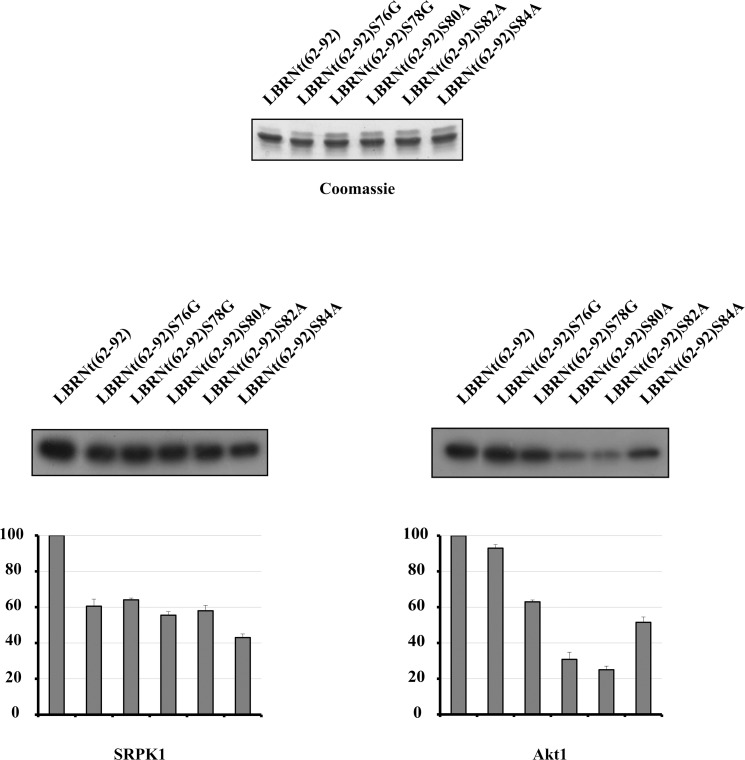
Fig 4. Determination of the sites phosphorylated by SRPK1 and Akt1.
Phosphorylation of GST-LBRNt(62–92), GST-LBRNt(62–92)S76G, GST-LBRNt(62–92)S78G, GST-LBRNt(62–92)S80A, GST-LBRNt(62–92)S82A και GST-LBRNt(62–92)S84A by 0.19 μM GST-SRPK1 (left panel) and 0.07 μM Akt1 (right panel). Only the relevant part of the autorad corresponding to the phosphorylated recombinant proteins is shown. Enzyme activity is expressed as a percent of the activity obtained with GST-LBRNt(62–92) which was set to 100 percent. Data represent the means ± SE of three independent experiments. On top of the figure we show a Coomassie Blue staining of the recombinant proteins (1.95 μM of each) used in the phosphorylation assays.

