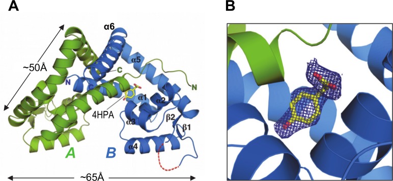
Fig 2. The crystal structure of NadR in complex with 4-HPA.
(A) The holo-NadR homodimer is depicted in green and blue for chains A and B respectively, while yellow sticks depict the 4-HPA ligand (labelled). For simplicity, secondary structure elements are labelled for chain B only. Red dashes show hypothetical positions of chain B residues 88–90 that were not modeled due to lack of electron density. (B) A zoom into the pocket occupied by 4-HPA shows that the ligand contacts both chains A and B; blue mesh shows electron density around 4-HPA calculated from a composite omit map (omitting 4-HPA), using phenix [38]. The map is contoured at 1σ and the figure was prepared with a density mesh carve factor of 1.7, using Pymol (www.pymol.org).

