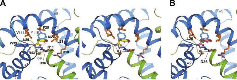
Fig 4. Atomic details of NadR/HPA interactions.
A) A stereo-view zoom into the binding pocket showing side chain sticks for all interactions between NadR and 4-HPA. Green and blue ribbons depict NadR chains A and B, respectively. 4-HPA is shown in yellow sticks, with oxygen atoms in red. A water molecule is shown by the red sphere. H-bonds up to 3.6Å are shown as dashed lines. The entire set of residues making H-bonds or non-bonded contacts with 4-HPA is as follows: SerA9, AsnA11, LeuB21, MetB22, PheB25, LeuB29, AspB36, TrpB39, ArgB43, ValB111 and TyrB115 (automated analysis performed using PDBsum [44] and verified manually). Residues AsnA11 and ArgB18 likely make indirect yet local contributions to ligand binding, mainly by stabilizing the position of AspB36. Bond distances for interacting polar atoms are provided in Table 3. Side chains mediating hydrophobic interactions are shown in orange. (B) A model was prepared to visualize putative interactions of 3Cl,4-HPA (pink) with NadR, revealing the potential for additional contacts (dashed lines) of the chloro moiety (green stick) with LeuB29 and AspB36.

