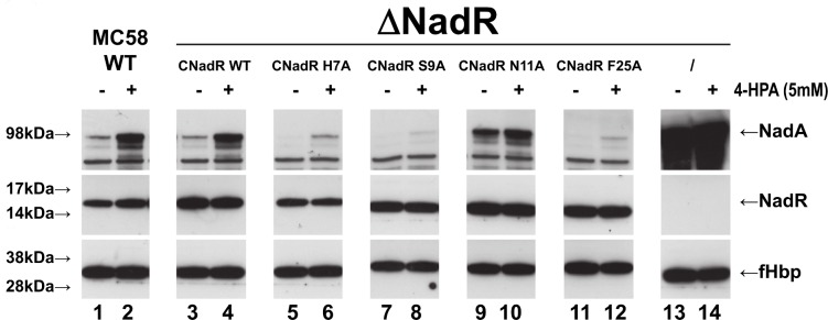
Fig 9. Structure-based point mutations shed light on ligand-induced regulation of NadR.
Western blot analyses of wild-type (WT) strain (lanes 1–2) or isogenic nadR knockout strains (ΔNadR) complemented to express the indicated NadR WT or mutant proteins (lanes 3–12) or not complemented (lanes 13–14), grown in the presence (even lanes) or absence (odd lanes) of 5mM 4-HPA, showing NadA and NadR expression. Complementation of ΔNadR with WT NadR enables induction of nadA expression by 4-HPA. The H7A, S9A and F25A mutants efficiently repress nadA expression but are less ligand-responsive than WT NadR. The N11A mutant does not efficiently repress nadA expression either in presence or absence of 4-HPA. (The protein abundance levels of the meningococcal factor H binding protein (fHbp) were used as a gel loading control).

