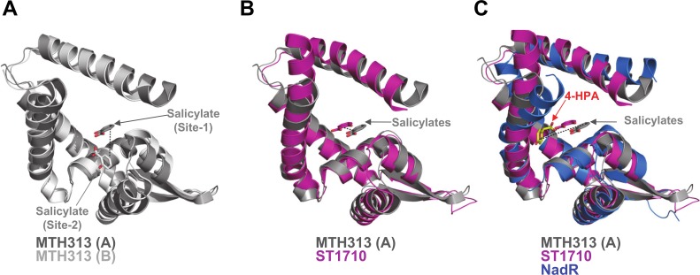
Fig 10. NadR shows a ligand binding site distinct from other MarR homologues.
(A) A structural alignment of MTH313 chains A and B shows that salicylate is bound in distinct locations in each monomer; site-1 (thought to be the biologically relevant site) and site-2 differ by ~7Å (indicated by black dotted line) and also by ligand orientation. (B) A structural alignment of MTH313 chain A and ST1710 (pink) (Cα rmsd 2.3Å), shows that they bind salicylate in equivalent sites (differing by only ~3Å) and with the same orientation. (C) Addition of holo-NadR (chain B, blue) to the alignment reveals that bound 4-HPA differs in position by > 10 Å compared to salicylate, and adopts a novel orientation.

