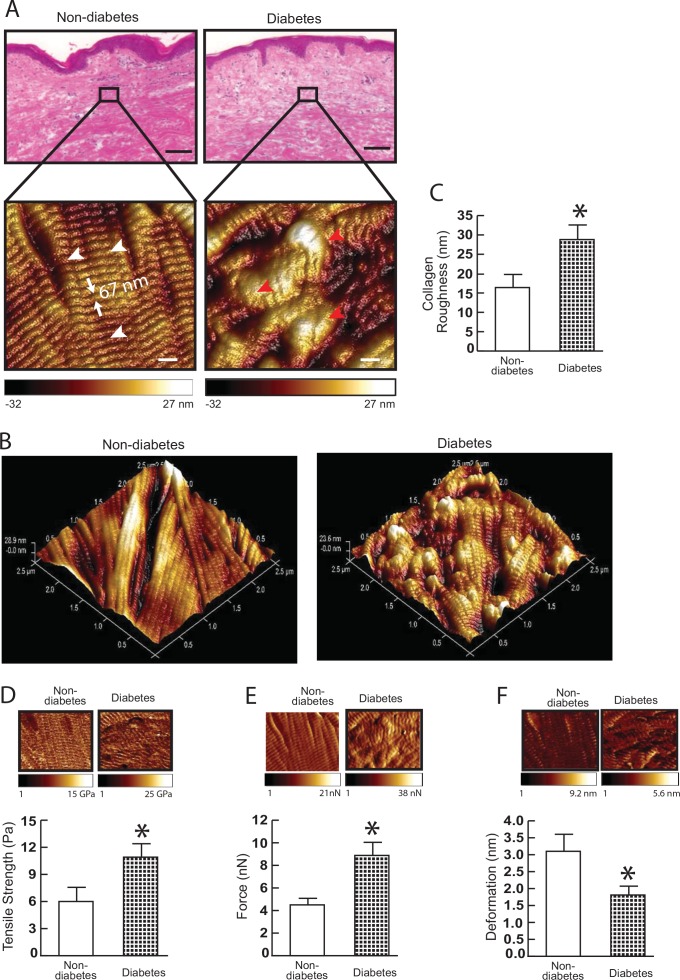
Fig 3. Collagen fibril nanoscale morphology and mechanical properties in diabetic skin.
(A) Nanoscale collagen fibrils were imaged by atomic force microscopy (AFM). The white and red arrow heads indicate intact and fragmented/disorganized collagen fibrils, respectively. Images are representative of nine subjects. (B) Three-dimensional nanoscale collagen fibrils were imaged by AFM. Images are representative of nine subjects. (C) Collagen fibril roughness was analyzed using Nanoscope Analysis software (Nanoscope_Analysis_v120R1sr3, Bruker-AXS, Santa Barbara, CA). Results are expressed as the mean ± SEM, N = 6, *p<0.05. (D) Tensile strength, (E) traction forces, and (F) deformation were determined by AFM PeakForceTM Quantitative NanoMechanics mode and Nanoscope Analysis software. Means±SEM. N = 8, *p<0.05.

