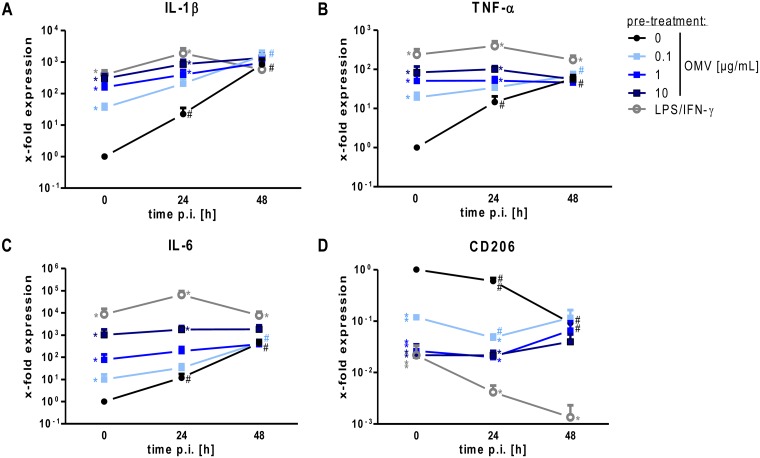
Fig 3. OMVs reduce responsiveness of macrophages to L. pneumophila infection.
(A-D) THP-1 cells treated as described in Fig 2A and RNA samples were taken at the time of infection (0 h) or 24 and 48 h post infection (p.i.). qPCR was performed for markers of classically activated macrophages (A: IL-1β, B: TNF-α, C: IL-6) and alternatively activated macrophages (D: CD206). Results are normalized to untreated control cells at every time point. Mean values of three independent experiments are shown. Statistics: Mann-Whitney test; * p<0.05 compared to 0 μg/mL OMVs. # p<0.05 compared to equally treated 0 h sample.

