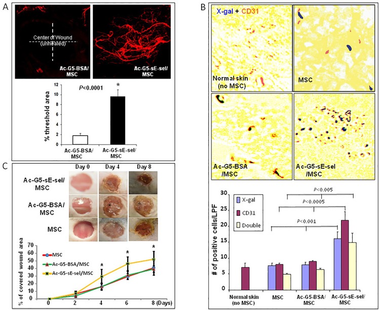
Fig 4. Local and systemic delivery of Ac-G5-sE-sel nanocarrier coated MSC to murine wound tissues.
A: Pro-angiogenic effect of locally injected Ac-G5-sE-sel nanocarrier coated MSC to wounded db/db mice. 1 x 10⁶ of MSC from donor db/db were coated with Ac-G5-sE-sel or Ac-G5-BSA nanocarriers, and directly injected into wound bed of wounded db/db mice. (top) Representative images of confocal microscopy of Dil perfusion; (bottom) quantitative data of Dil-stained blood vessel density in wound (n = 6 mice/group). B: Homing of systemically delivered Ac-G5-sE-sel nanocarrier coated MSC to wound tissues. 1 x 10⁶ of MSC (LacZ+/Luc2+) from donor mice were coated with Ac-G5-sE-sel or Ac-G5-BSA nanocarriers, and systemically infused into wounded C57BL/6 mice. (top) Representative images of β-gal and CD31 staining (brown) of wound and normal skin tissues. IHC was performed on harvested wounds at day 8 post cell administration in frozen samples that were also subjected to β-gal (blue) staining. Normal skin tissues were used as negative control. (bottom) Quantitative data of β-gal+, CD31+ (single positive) and β-gal+/CD31+ (double positive) cells (X10). Numbers of positive cells were counted from 5 randomly selected fields in wound and skin samples. Data are percentage of mean ± SD (n = 6 mice/group). C. Pro-healing effect of systemic delivery of Ac-G5-sE-sel coated MSC. Healing rate expressed as percent of wound re-epithelialization (covered with new skin). Top: representative wounds at different days are shown for each group. Bottom: wound healing rate. Data are percentage of mean ± SD (n = 6 mice/group).

