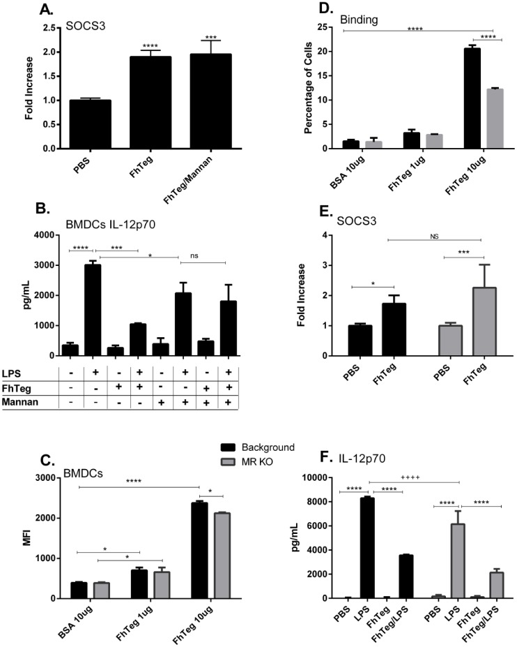
Fig 5. The immune properties of FhTeg are independent of the MR receptor.
A: BMDCs were stimulated with mannan for 30 min prior to incubation with FhTeg for 2.5 h. Total RNA was extracted, and after reverse transcription cDNA was analyzed with qPCR for SOCS3. RNA expression was normalized to GAPDH and actin control genes. B: BMDCs were pre-incubated with mannan prior stimulation with PBS or FhTeg (10μg) before addition of LPS (100ng ml-1) for 18 h. IL12p70 levels were measured with commercial ELISA kits. Data are presented as the mean ± SEM of two independent experiments. ***p ≤ 0.001; ****p ≤ 0.0001 compared to LPS group. C,D: BMDCs isolated from MR-knockout mice were stimulated with fluorescently labelled FhTeg or BSA (10μg ml-1, green) for 45 min prior to paraformaldehyde fixation. FhTeg binding to cells was assessed by flow cytometry and reported in bar chart format. E: BMDCs isolated from MR-knockout mice were stimulated with FhTeg (10μg) for 2.5 h. Total RNA was extracted, and after reverse transcription cDNA was analyzed with qPCR for SOCS3. RNA expression was normalized to GAPDH and actin control genes. F. BMDCs derived from MR knockout mice were stimulated with PBS or FhTeg (10μg) before addition of LPS (100ng ml-1) for 18 h. IL12p70 levels were measured with commercial ELISA kits. Data are presented as the mean ± SEM of two independent experiments. *p ≤ 0.05, **, p ≤ 0.01; ***, p ≤ 0.001.

