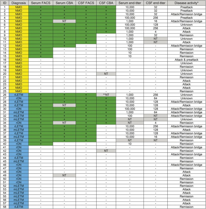
Figure 2. Concordance of results for commercial cell-based assay (CBA) and fluorescence-activated cell sorting (FACS) assays, FACS titers, and disease activity at draw date in time-matched paired specimens of serum and CSF from patients with neuromyelitis optica (NMO) and high-risk (HR) NMO (figure 3B plots serum endpoint vs CSF endpoint in these time-matched paired samples).
Positive samples are marked with a green background. Samples not tested (NT) are marked with a gray background. Patients with NMO are highlighted in yellow; HR-NMO patients in blue. mLETM = monophasic longitudinally extensive transverse myelitis; rLETM = recurrent longitudinally extensive transverse myelitis; rON = recurrent optic neuritis.

