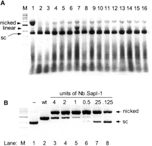
Figure 3.

(A) Initial isolation of Nb.SapI-1 (variant 33). Nicking activity is revealed by incubating cell extract with supercoiled pUC19; (sc), supercoiled. Lane M is a 1 kb DNA ladder where the prominent band is a 3 kb band. The nicked form of pUC19 migrates at >3 kb. Lane 1 contains a significant level of nicked pUC19 produced by variant 33 extract. Lanes 2–16 (variants 34–48) contain typical levels of nicked pUC19 produced by the non-specific nicking activity of E.coli extract. Variant 39 (lane 7) displays double-stranded cleavage activity as linearized pUC19 (2686 bp) is observed. (B) DNA nicking activity of purified Nb.SapI-1 (variant 33). Lane 1 is supercoiled pUC19 without enzyme addition (−). Lane 2 is pUC19 linearized by wt SapI. Lanes 3–8 were incubated with 4, 2, 1, 0.5, 0.25 and 0.125 U of Nb.SapI-1 (variant 33) for 60 min at 37°C, respectively.
