
Figure 1.
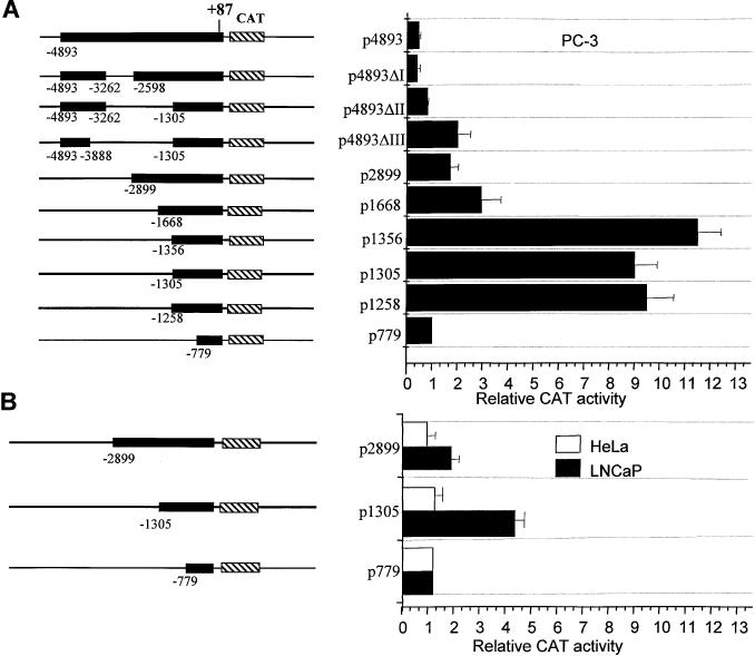
The effect of deletion in the 5′ region of the PAcP promoter on the CAT activity in human prostate cancer cells. (A) PC-3 cells and (B) LNCaP and HeLa cells. On the left, a schematic representation of the 5 kb PAcP promoter and its deleted variants inserted upstream of the CAT gene in the reporter plasmid pCAT-Basic. The numbers indicate the 5′ and the 3′ ends of the promoter DNA inserts, in relation to the transcription start site (+1). Cells were transfected with 1 μg of the indicated PAcP-CAT reporter constructs and assayed for CAT activity as described under Materials and Methods. On the right, the CAT activity is presented as the ratio of the tested construct to p779. Values were normalized for transfection efficiency by co-transfection with the β-galactosidase expression plasmid. Bars represent the SE of triplicates from at least two sets of independent experiments (n ≥ 6).