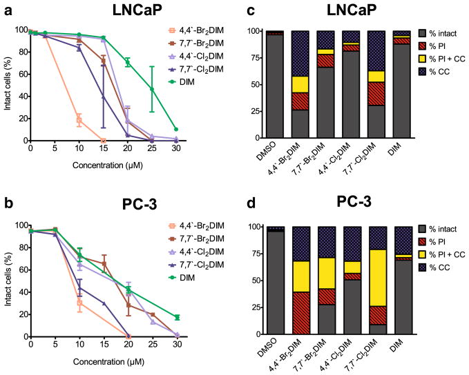
Fig. 2.
Ring-DIMs kill AD and AI prostate cancer cells via apoptotic and necrotic forms of cell death. The percentage of intact LNCaP cells (a) and PC-3 cells (b) that do not have fragmented nuclei, condensed chromatin, or propidium iodide staining was calculated 48 h after treatment with increasing concentrations (5–30 μM) of 4-4′-dibromo-DIM (□), 7-7′-dibromo-DIM (■), 4,4′-dichloroDIM (△), 7,7′-dichloroDIM (▲) or DIM (●). Fragmented nuclei and condensed chromatin are indicative of apoptosis, whereas propidium iodide staining is indicative of plasma membrane permeabilization and necrosis. Percentages are represented as mean ± SEM (n=3–5). (c) The percentage of LNCaP cells showing condensed chromatin (CC), propidium iodide staining (PI), or both (CC+PI) after a 48 h treatment with either 10 μM 4,4′-dibromoDIM, 15 μM 7,7′-dibromoDIM, 15 μM 4,4′-dichloroDIM, 15 μM 7,7′-dichloroDIM or 15 μM DIM. (D) The percentage of PC-3 cells showing CC, PI, or CC+PI after a 48 hourtreatment with either 20 μM of the ring-DIMs or DIM

