Fig. 4.
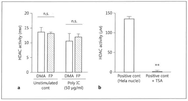
HDAC activity in FP-treated BEAS-2B cells, a BEAS-2B cells were stimulated with poly IC (50 μg/ml) in combination with either FP (10−7 M) or DMA for 24 h. Nuclear extracts were then prepared and assayed for HDAC activity, b Nuclei isolated from HeLa cells were either untreated or incubated with 100 nM of the HDAC inhibitor TSA for 30 min. Nuclear extracts were then assayed for HDAC activity. The data are the mean ± SEM of 3 independent experiments. ** p < 0.05 compared with positive control HeLa nuclei without TSA. cont = Control; n.s. = not significant.

