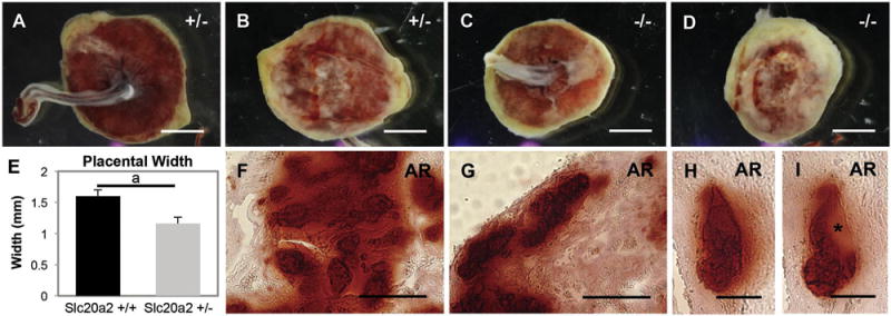
Fig. 5.

Placental calcification is abundant in Slc20a2 deficient placentas. Placentas from Slc20a2+/− females were analyzed. Brightfield whole mount images suggest abnormal vascular development in placentas from Slc20a2−/− embryos compared to those from Slc20a2+/− embryos (A–D). Placentas from Slc20a2+/− females were thinner than control placentas (p = 0.006) (E). Positive staining for Alizarin Red was seen in the vascularized labyrinth (F), and the spiral arteries of the decidua (G–I) indicating calcification in these areas. Consecutive sections of calcified spiral arteries revealed nodules of basement membrane-like material highlighted by an asterisk (H, I). Scale bars: A–D = 1 μm, F–G = 50 mm, H–I = 25 μm. ap < 0.05.
