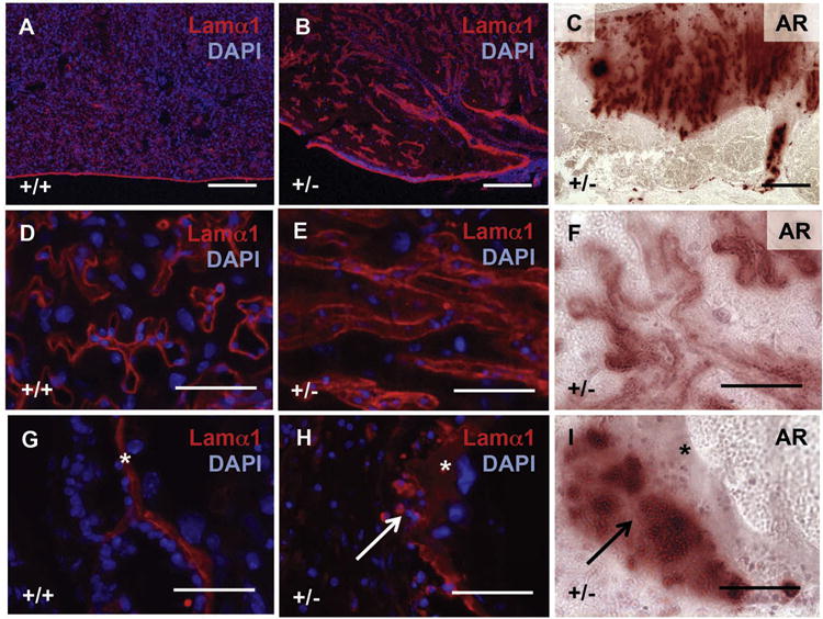
Fig. 8.

Abnormal vasculature associates with placental calcification in Slc20a2 deficient placentas. Detection of Lamα1 with DAPI nuclear counterstaining reveals abnormal vascular structure to the labyrinth (A, B and D, E), as well as thickening of the chorioallantoic plate (G, H). Alizarin Red staining of consecutive sections reveals close proximity of calcified sites to Lamα1 in Slc20a2+/− placentas. Scale bars: A–C = 300 μm, D–I = 50um. Asterisks: basement membrane. Arrows: calcified sites.
