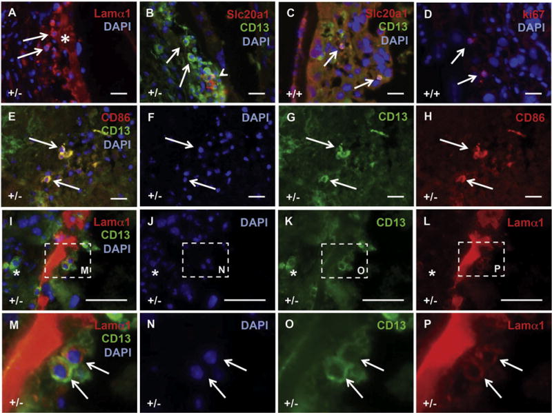
Fig. 9.

A novel Lamα1 and CD13 positive placental cell is present at the calcified sites. Mouse placental tissue from Slc20a2 +/− and Slc20a2+/+ females was stained with a variety of antibodies to characterize the cellular milieu at calcified sites, and to determine the cellular identity of candidate causative cell types. Cells expressing Lamα1 (arrow) were identified adjacent to the enlarged basement membranes (asterisk) in calcified lesions (A). CD13 positive (arrows) and CD13/Slc20a1 positive cells (arrow head) were detected in the calcified lesions (B). Morphologically, similar cells that co-expressed CD13 and Slc20a1 were sparse in WT placenta. Staining of consecutive sections revealed expression of ki67 (D). Round CD13 positive cells in the labyrinth are CD86 positive (E–H). CD13 positive cells at the calcified site adjacent to enlarged basement membranes contain Lamα1 (arrows), while other CD13 positive cells do not (asterisk) (I–P). Scale bars: A–L = 20 μm. M–P: magnified images as indicated by dashed lines in I–L.
