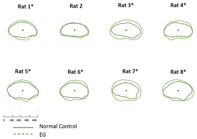
Figure 3. EG (dotted line) versus Control (solid line) eye Optic Nerve within the Anterior Scleral Canal Opening (ON-ASCO) comparison for each study animal.
ON-ASCO data points for the Control and EG eye of each rat are schematically overlaid using the ON-ASCO centroid of each eye. All data are in right eye orientation. The scale is in micrometers. (*) denotes animals in which Global EG versus Control Eye ON-ASCO radius differences achieved significance (Supplemental Table 2).

